

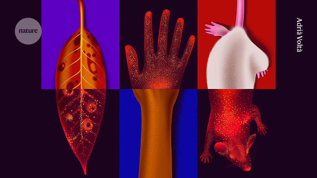
In 2025, quantum physicist Daniel Oblak and his colleagues published images of mice that made headlines around the world. The researchers put four anaesthetized mice inside a dark chamber and pointed a sensitive camera at them. It picked up a stream of photons emitted from the animals’ skin: a light signal too faint to be seen by the human eye, but visible in the camera images as a ghostly, mouse-shaped glow1.
The mice were killed at the end of the experiment, and almost as an afterthought, the team placed the dead animals back into the chamber to image them again. This time, the glow had disappeared; aside from a few remaining spots of light, the mice had essentially dissolved into background noise. Seeing the images was “a defining moment”, says Oblak, who is based at the University of Calgary in Canada. It was as if the researchers had photographed life itself.
A sensitive camera picks up biophoton emissions from a mouse (alive, left; dead, right).Credit: Lana Frankle/University of Calgary
The study went viral. The researchers were inundated with messages, from a healer’s claim that his hands emitted light, to eyewitness reports of glowing trees; headlines around the world asked whether auras are real. Oblak emphasizes that the photons he captured have little to do with such claims, not least because the naked eye can’t see the faint emissions. He sees it instead as the mark of a long-misunderstood field finally coming of age: the study of the ‘biophotons’ produced by living cells across the tree of life.
That biophotons (also known as ultraweak photon emissions) exist had been established long before Oblak’s team took the striking images. The emissions stem from metabolic processes inside cells, typically in their mitochondria. But he and other researchers are studying them in the hope that they will be useful as subtle markers of disease or health. And if scientists can confirm hints that cells themselves also react to the photons, that could expand their understanding of how life works. “If you can prove that cells communicate by light, it would be fantastic,” says Michal Cifra, a specialist in bioelectrodynamics at the Czech Academy of Sciences in Prague.
“It’s one of those fields that feels like it is on the edge of respectability,” says Nick Lane, who studies mitochondrial biochemistry at University College London. “That doesn’t mean it’s wrong.”
A century of study
The idea that cells might communicate through extremely low-intensity light was first suggested in the 1920s by Russian biologist Alexander Gurwitsch2. He conducted experiments with onion roots, reporting that a growing root tip could trigger cell division in a neighbouring root. The effect was blocked if the roots were separated by opaque or glass plates, but persisted if the plate was made of quartz, which lets through ultraviolet radiation. He concluded that the root cells were producing — and responding to — photons of UV light. A flurry of research followed, says Cifra, but the effect was hard to replicate. “People lost interest and thought it was just pseudoscience.”
In the 1950s, studies with photomultiplier tubes, devices that convert faint light into electric signals and are sensitive enough to count individual photons, confirmed that living systems, from bacteria to plants and mammals, do indeed emit light3. Its intensity falls slightly below the limit of dark-adapted conscious human vision, at just tens to hundreds of photons per square centimetre per second, and it covers a range of wavelengths that include visible light, from UV to near-infrared2. This phenomenon is distinct from both thermal radiation (which is produced by all surfaces, living or not, as a function of temperature) and the much brighter light produced by bioluminescent organisms, such as fireflies and jellyfish.
Can flashing lights stall Alzheimer’s? What the science shows
But the field was unable to shake off connotations of pseudoscience. From the 1970s to the early 2000s, German biophysicist Fritz-Albert Popp led a body of research based around the controversial claim that biophotons (a term that he coined) aren’t emitted randomly but are structured more like laser light, organized by a coherent quantum electromagnetic field that permeates the organism or cell. This, together with the fact that Popp and his supporters suggested biophotons could be a mechanism for the effects of treatments from homeopathy to acupuncture, caused many mainstream researchers to avoid the topic.
There is not yet evidence for such quantum coherence, says Cifra, adding that it’s hard to imagine how it might arise in a biological system. But that doesn’t mean the emissions aren’t worth studying, he says. Like Oblak, he hopes that with more sensitive detectors, combined with rigorous methods, biophotons could become a more established area of inquiry. “I would say the field, the technology and also the mindset of certain parts of the community has matured.”
Light and health
Oblak’s life-versus-death mouse study was a striking visual demonstration of how the photons are intimately linked to life: “It was the first time you could really see it side by side,” says Oblak’s colleague at the University of Calgary, Christoph Simon, who is a theoretical quantum physicist. But the hope is that biophotons could provide a window into the health of a cell.
Biophotons are produced during aerobic metabolism, in which cells use oxygen to break down nutrients and extract energy. This mostly occurs in mitochondria3. During metabolic reactions, short-lived chemicals produced from oxygen, called reactive oxygen species (ROS), generate intermediates that decompose to produce molecules in an ‘excited’ electronic state. As these relax into more stable arrangements, they can release energy in the form of photons. A high-energy ‘singlet’ state of oxygen can emit a photon of red light as it decays, for example, whereas excited carbonyl (C=O) groups glow in the blue-green range.
Although they are a part of normal metabolism, ROS also function as signalling molecules when a cell responds to stress, such as infection or injury. And many studies — looking at a range of organisms, from amoebas to human hands — have shown that these stress responses accordingly cause a burst of emitted light2. In unpublished experiments, Oblak and Simon made small cuts in leaves from a dwarf umbrella tree (Heptapleurum arboricola). They immediately saw a burst in photon emission at the cut site, which soon spread to form a wider ring; Oblak suggests that they were watching the plant’s response to the injury evolving in real time. The team also detected changing patterns in the wavelength of the emitted light. “The stress response seems to be different than the purely metabolic response of something growing,” Oblak says.

Biophoton emissions from a cut leaf change over three days.Credit: Vishnu Seshan/University of Calgary
“I’m fascinated by the fact that just by watching the light, one can potentially assess the liveliness, the metabolic activity, the degree of oxidative stress,” says Cifra. Cancer, cardiovascular disease and neurodegenerative diseases are all associated with increases in oxidative stress, he points out, so imaging biophotons might enable early detection of such diseases. “This is very exciting.”
Several teams are already investigating whether biophotons can reveal early signs of cancer. Earlier this year, a team led by Maurizio Benfatto, a photonics researcher at Italy’s National Institute of Nuclear Physics in Frascati, studied emissions in brain cells. The team reported differing patterns of emissions in healthy cells called astrocytes compared with cancerous cells called glioblastoma cells4. Nirosha Murugan, a biophysicist at Wilfrid Laurier University in Waterloo, Ontario, and her colleagues have found the same for healthy skin cells versus cancerous melanoma cells, in mouse and human cell lines5. She is now recruiting participants to test whether photon counts can discriminate between benign moles and melanomas on people’s skin, without needing to take a biopsy.
Researchers don’t necessarily need state-of-the-art equipment to look at biophotons — depending on what they’re trying to capture. Oblak’s team used a sensitive CCD (charge-coupled device) camera, cooled to nearly −100 °C, to take detailed images of mice, but Murugan’s work, which aims only to count photons, uses simple photomultiplier tubes pointed towards people who are completely shielded from light.

The University of Calgary team: Daniel Oblak, Vishnu Seshan, Vahid Salari, Christoph Simon.Credit: Riley Brandt/University of Calgary
Because cancer cells have different metabolic states from non-cancer cells, “it’s not too surprising that there are differences” in biophoton emissions, says Brian Wilson, a medical biophysicist at the University of Toronto, Canada. He suggests that biophotons might therefore be useful as a research tool, perhaps for screening how drugs affect cells. But he’s not convinced they could reveal useful information from patients. “I have a hard time seeing a clinical diagnostic,” he says. “The signal is just far too weak.”
The original aim of Oblak’s mouse study was to test whether biophotons could reveal the presence of tumours under the animals’ skin, but there was no conclusive signal in the images, he says, possibly because any excess photons were scattered by dead tissue around the tumours, or by the skin itself.
The team has, however, reported that biophoton emissions from hippocampus tissue taken from healthy rats differ from those produced by tissue taken from rats with a chemically induced form of Alzheimer’s disease6. The research is at an early stage, but the team suggests that, in future, it might be possible to monitor for signs of the disease with a tiny photonic chip attached to the inside of the skull. Other uses of biophotonics being investigated include scanning plant seeds for viability and checking the health of donated organs before they are transplanted. “It’s a whole new signal you can look at,” says Simon. “I think we have really only scratched the surface.”
Cellular communication?
These studies raise the question of whether living systems are reading or reacting to these signals, as Gurwitsch suggested originally with the onion roots. Do biophotons represent a previously unrecognized cellular communication system? This suggestion is much more controversial than the idea of using them for diagnostic purposes.
Numerous studies have reported that cells separated by transparent barriers can influence each other, from activating white blood cells to adjusting calcium signalling or growth rate2. Cifra, who has written several critical reviews of the field7, says that these effects are often hard to reproduce, and that the studies don’t always adequately rule out other causes, such as chemicals being transferred through the air.

Forget SkinTok: the real science of skincare and why it matters for your healt
Some experiments are harder to dismiss, says Cifra. A team led by biologist Rhys Mould at the University of Westminster, London, for example, studied isolated mitochondria in sealed quartz containers. Adding a toxin to one container decreased the respiration rate of mitochondria in adjacent ones, and mitochondria from cancer cells showed a stronger response than those from healthy cell lines did. The effect was blocked by an opaque barrier of aluminium foil8. Even so, Cifra argues that it’s difficult to see how biophoton signalling could work without background light swamping the weak emissions. “I’m not discarding the possibility for this phenomenon to be real,” he says. But if it is, “we are probably missing something important in the underlying physics”.
One feature that could help the emissions to stand out from background noise is if the signal is in the overall, coordinated pattern of emissions, rather than in each individual photon. Benfatto says that he found such higher-level patterns when he looked at how photon emissions from germinating lentil seeds vary over time9. This doesn’t prove that other plants are detecting the photons but, if confirmed, it hints that biophotons could at least carry useable information. Benfatto suggests this might enable different seeds or plants in the soil to influence each other’s growth patterns, or trigger germination.
Researchers are also examining whether biophotons might be sensed by cells inside our bodies. Benfatto has reported preliminary evidence of complex patterns in biophoton emissions from astrocytes,which seem to be weaker in cancerous cells4. Again, this might be only a by-product of altered metabolism, and not in itself an active driver of cellular health or disease. But if the photons do have a role in regulation, he suggests, understanding them might lead not just to tests but also to therapies, which could help to retune the system when it goes wrong. “Perhaps this can be done with light.”
The surprising science behind red-light therapy — and how it really works
Indeed, some researchers have suggested that biophoton signalling might be a mechanism by which red-light (or near-infrared-light) therapies seem to influence many biological processes, from skin ageing and joint pain to neurodegenerative disorders (see Nature 651, 871–874; 2026).
